Nobel prize winning virologist Harald zur Hausen sent our scientific director Dharam Ablashi the attached electron microscope image of HHV-6A (GS) virions that were propagated in an HSB2 cell line and would like help in identifying the multiple small particles attached to the viral envelope shown in the photograph below. Zur Hausen reports that the particles did not react to HHV-6 or any other monoclonal antibodies. The particles appear to have diameters of about 40 nm.
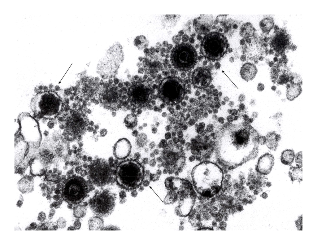
Do you have any thoughts on what they might be?
Yasuko Mori and Koichi Yamanishi published a paper in 2008 indicating that HHV-6 acquires its envelope via a multivesicular body or exosome pathway (Mori 2008). Could these small particles be exosomes?
Another possibility suggested by HHV-6 expert Philip Pellet, PhD from Wayne State University is that another virus could be present in the culture, such as a bovine viral diarrhea virus (approximately 50 nm compared to 200 nm for HHV-6).
What are your thoughts? Please email Dharam Ablashi.