
Could HHV-6B infection exacerbate mitochondrial disease?
The increasing speed and decreasing cost of rapid sequencing is facilitating a better understanding of the human virome—its diversity, organ-specificity and interactions with host cells. A team led by the University of Helsinki analyzed the variability and integration sites of nine persisting viruses in nine different organs (96 autopsy samples) from thirteen individuals who died suddenly from non-viral causes (mainly trauma and heart disease). The organs included brain, colon, heart, liver, lung, kidney, skin, whole blood and plucked hair. The viruses studied included herpesviruses 1, 3, 4, 5, 6B and 7, parvovirus B19, Merkel cell polyomavirus (MCPyV) and JC polyomavirus (JCPyV).
The team analyzed intra-host, cross-organ variability by comparing the viral consensus sequences reconstructed from different organs in an individual. In addition, they determined within-organ variability by conducting minor variant (MV) analysis of non-consensus variants. In addition, the investigators explored viral integrations into the host DNA by assessing chimeric (viral-host) sequencing reads.
The analysis unexpectedly revealed integrations of HHV-6B into mitochondrial DNA. HHV-6B integrations into mitochondrial DNA occurred in three kidney samples. One of these junctions was between the unique region (U36 gene) of HHV-6B and the mtDNA NADH dehydrogenase subunit 5 (MT-ND5). This is interesting given the in vitro studies indicating that HHV-6 infection can disrupt mitochondrial anatomy and function (Schreiner 2020; Prusty 2018). The Schreiner 2020 study demonstrated how iciHHV6 cells can be stimulated to secrete factors that simultaneously cause mitochondrial fragmentation, a reduction in intracellular ATP reserve and an expanded antiviral defense by their neighboring cells. HHV-6 encephalitis can trigger fatal disease in children with underlying POLG mitochondrial disorders (Al-Zubeidi 2014).
Other conclusions of the Finnish study were:
- Non-clonal viral integrations into host DNA occur even in individuals without cancer. The analysis identified 47 integrations for 6 viruses (HHV -6B, HHV -7, B19V , MCPyV , EBV and HSV-1) from 127 genomes, backed by, on average, eight supporting reads and four split reads per junction. As would be expected, most integrations occurred with HHV-6B: in total, 33 integrations across 7 individuals and 12 samples were identified. The study also identified MCPyV integrations and truncations resembling clonally expanded variants in Merkel cell carcinomas.
- The viral sequences across organs were remarkably conserved within each individual, suggesting that persistence stems from single dominant strains. Since the autopsy specimens examined came from immunocompetent individuals who died of non-viral causes, the team concludes that intra-host viral evolution is unusual in such people. In immunocompromised individuals, other studies have found greater intra-host variation of individual viruses. In addition, the Helsinki team detected increased sequence diversity in the two individuals (out of 13 studied) with viral reactivations, suggesting that replication status influences diversity. The data are summarized in Figure 1.
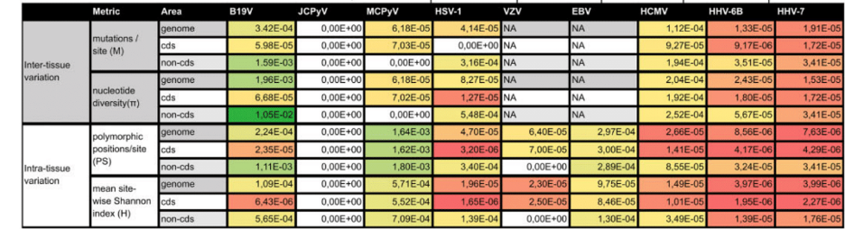
Figure 1. Exact values of all the metrics of diversity (mutation / site, nucleotide diversity, polymorphic positions / site and mean site-wise Shannon index). Higher values are in shades of green and lower in yellow and red. B19V, human parvovirus B19; EBV, Epstein-Barr virus; HCMV, human cytomegalovirus; HHV-6B , human herpesvirus 6B , HHV-7, human herpesvirus 7; HSV-1, herpes simplex virus 1; JCPyV, JC polyoma virus; MCPyV, Merkel cell polyoma virus; VZV, varicella-zoster virus.
- Both HHV-6B and HHV-7 exhibited lower intra-host diversity across all four metrics compared to other viruses studied. The exception was JCPyV, which showed no identifiable intra-host variability.
- Intra-organ diversities were minimal: on the order of 10-6 for HHV-6B and HHV-7, two magnitudes higher for parvovirus B19, and three magnitudes higher for MCPyV.
For investigators interested in HHV-6 and HHV-7, this study adds to a literature indicating that infection with these viruses can affect a cell’s mitochondria and energy-producing capacity, and indicates the need for further study of this question.
Read the full text: Pyoria 2024