Investigators at the National Cancer Institute found that HHV-6B can be identified in lymphoid pathologies including Hodgkin lymphoma (HL) and angioimmunoblastic T-cell lymphoma (AITL) in addition to lymphadenitis. They warn that failing to identify HHV-6 can lead to misdiagnosis of benign and malignant lymphoproliferative conditions.
Pathology investigators at the Center for Cancer Research at NCI/NIH evaluated lymph nodes from neoplastic and reactive lymphadenitis cases positive for HHV-6. Specimens were obtained from several cases of reactive lymphadenitis and lymphoma (AITL and one classic HL).
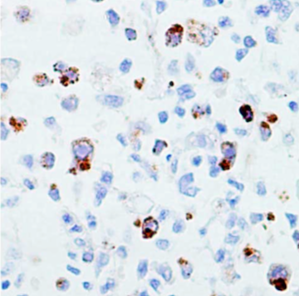
HHV-6-infected cells with membranous and Golgi pattern.
Source: Jayalakshmi 2018, American Journal of Surgical Pathology
They found that the lymphadenitis cases all showed complete or partial effacement of architecture with marked paracortical hyperplasia. In addition, 4/5 lymphadenitis cases showed numerous large atypical lymphoid cells, with pleomorphic nuclei, vesicular chromatin and prominent eosinophilic intranuclear inclusions. The inclusions were limited to CD4+ cells.
The authors conclude that failure to note the presence of HHV-6 in reactive lymph nodes could result in misinterpretation of a benign lymphoproliferative disorder as a malignant condition, particularly peripheral T-cell lymphoma. Also, the HHV-6-associated presence of large atypical cells resembling immunoblasts with prominent eosinophilic intranuclear inclusions could suggest classic HL.
Finally, they note that the presence of HHV-6-induced neutrophilic infiltrates and necrosis in cases of AITL and HL may also obscure the morphology of the underlying lymphoma, making identification of the lymphoma subtype more difficult.
The cases of AITL lacking viral inclusions were all negative for HHV-6 by immunohistochemistry, and the staining for HHV-6 was negative in the Reed Sternberg cells of the HL case. The authors used only a late protein and did not stain with a latent protein or HHV-6 DR6/7 antibodies described by others (Lacroix 2010, Siddon 2012). A recent study found HHV-6 in 86% of nodular sclerosis HL cases and in the Reed-Sternberg cells in nearly half of those cases (Siddon 2012).
HHV-6 immunohistochemistry is rarely performed in lymphadenitis biopsies and is not listed as a possible cause in most reviews on the subject. In addition to avoiding mistakes in the diagnosis of malignant conditions, HHV-6 IHC would also benefit patients with undiagnosed viral lymphadenitis.
Read the full paper: Balakrishna 2018.