

Virus in serum free medium on the left has a clean surface, while virus incubated in serum for an hour has accumulated protein corona. Source: Nature Communications, Ezzat 2019.
Swedish researchers report that viruses interact with proteins to form a coating, or protein corona, that facilitates the formation of amyloid plaque, supporting previous findings by Harvard’s Rudy Tanzi and Robert Moir.
The first author Kariem Ezzat of Stockholm University and Karonlinska Institute explained, “Imagine a tennis ball falling into a bowl of milk and cereal. The ball is immediately covered by the sticky particles in the mix and they remain on the ball when you take it out of the bowl. The same thing happens when a virus gets in contact with blood or lung fluids that contain thousands of proteins. Many of these proteins immediately stick to the viral surface, forming a so-called protein corona.”
The investigators demonstrated that first viruses bind to amyloid proteins, and then the protein sheath sprouts amyloid fibrils which radiate from the virus. Further, they found that infecting mice with HSV1 accelerated the transformation of soluble amyloid proteins into the thread-like structures that make up amyloid plaques.
The Alzheimer’s model mice infected with HSV1 started showing signs of disease within 48 hours of infection, whereas without HSV1, the mice took several months to develop significant pathology.
In July 2018, Robert Moir and Rudy Tanzi demonstrated that HSV1, HHV-6A, and HHV-6B could each seed amyloid formation in their 3D cell culture model (Eimer 2018). Previously, Ruth Itzhaki’s lab showed that HSV1 causes beta-amyloid accumulation in cultured neuronal and glial cells (Wozniak 2007).
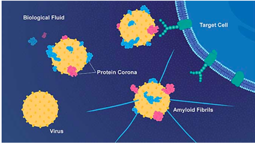
Virus developing a protein corona and fibrils.
Source: E. Wikander/Azote/Stockholm University
In a comprehensive mutiscale analysis led by Joel Dudly at Mt. Sinai, 515 viruses were studied but only HHV-6A and HHV-7 were found to have signifcantly more DNA and RNA in AD brains vs. controls (Readhead 2018). They also found regulatory relationships linking viral abundance and modulators of metabolism consistent with Alzheimer's.
The only other study to look at viruses in brain tissue was Itzhaki’s group, who looked for DNA using nested PCR. They found HHV-6 in 70% of AD brains compared to 40% of controls (p=0.003), while HSV1 was prevalent in both AD brains and controls (Lin 2002).
Read the full paper: Ezzat 2019.
An excellent summary of this paper can also be found on the Alzforum web site.