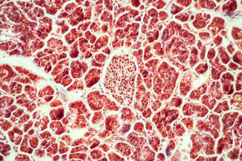
Investigators from the La Jolla Institute of Immunology used high-resolution confocal microscopy to detect high levels of HHV-6 protein in the pancreatic islet cells from donors with type 1 diabetes.

Matthias von Herrath, M.D, Professor, Division of Developmental Immunology, La Jolla Institute for Immunology
The first to identify HHV-6 in single islet cells, the investigators used both immunofluorescence to detect HHV-6 glycoprotein B (gB) and PCR to detect HHV-6 DNA. Image analysis quantified gB expression and found that islets from donors with type 1 diabetes (T1D) expressed higher levels of gB compared to non-diabetic donors. Furthermore, a similar increase in the expression of the HHV-6A/B gene, U67, as well as the detection of the HHV-6B receptor OX40 helped confirm active HHV-6B infection in these islets.
Single islet gB expression was correlated with neither the expression of MHC class 1 proteins nor CD8+ T cell infiltration which could have suggested a more direct connection between HHV-6 and T1D pathology. However, the other findings indicate HHV-6 may have an indirect role in T1D and its potential complications. For example, T1D may make pancreatic islets more susceptible to infections such as HHV-6 which can lead to increased gB expression which may lead to the development of pancreatitis.
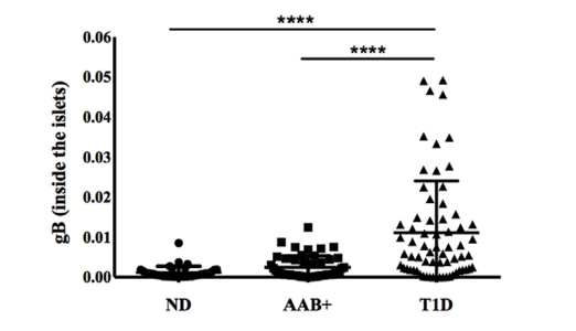
HHV-6 gB inside the islets of non-diabetic (ND), auto-antibody positive donors (AAB+) and Type 1 diabetes donors (T1D). Source: Journal of Autoimmunity, December 2019
Both enterovirus and herpesviruses have long been suspected of playing a role in T1D, but there has been little evidence from studies of the human pancreas to confirm a causal role of viruses in disease pathogenesis (Rodriquez-Calvo, 2016).
Based on their results, the authors call for further studies to understand how viral infections might trigger or facilitate the development of the disease.
Read the full paper: Sabouri 2019