Argument rests on the use of a putatively more sensitive diagnostic technique.
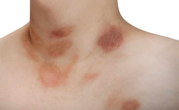
Pityriasis rosea (PR) is a skin condition of sudden onset that usually disappears over several weeks. First, a rose-colored “herald patch” with scaling borders appears, usually on the trunk. This is followed 1-2 weeks later by multiple small oval-shaped lesions on the trunk and limbs that often are very itchy. It long has been assumed that PR is likely caused by viral infection, but evidence of that has been relatively weak. However, over the past 5-10 years, reports from multiple laboratories have suggested that both HHV-6 and HHV-7 are linked to PR (Drago 2021; Drago 2015). Pityriasis rosea cases also increased following COIVD-19 or COVID vaccination (Wong 2023) leading some dermatologists to conclude that this could be due to HHV-6 reactivation (Dursun 2020).
A multi-institutional team from Turkey obtained skin biopsies and plasma samples from 25 patients with PR. Plasma also was obtained from 10 healthy control subjects.
The skin biopsies were obtained from both the initial herald patch and the subsequent oval lesions. IgG and IgM antibodies against HHV-6 and HHV-7 were tested by enzyme-linked immunosorbent assay and indirect immunofluorescence. Antibodies to EBV, CMV and parvovirus B19 also were measured.
A highly sensitive nucleic acid detection technique—calibrated quantitative RT-PCR (CQ RT-PCR)–was used to detect viral nucleic acid in skin: the investigators state that the sensitivity of the HHV-6 and HHV-7 kits was 0.82 copies of viral genome/µL and 0.4 copies of viral genome/µL, respectively. Viral loads in plasma were determined using REALQUALITY RS-HHV 6 (Code RQ-15; AB ANALITICA, Padova, Italy) and REALQUALITY RS-HHV 7 (Code RQ-19; AB ANALITICA, Padova, Italy) kits.
Seven skin biopsies from six (24%) patients detected HHV-6 or HHV-7 DNA. HHV-6 DNA positivity was obtained in ten patient plasma samples (with a range of 3.39–17.75 viral genome copies/µL). The two patients with the highest viral loads also had positive nucleic acid detected in the skin biopsies. HHV-6 DNA was positive in all the samples of one patient. However, five (50%) HHV-6 DNA positive (range 3.08–16.31 copies/µL) samples also were detected in the healthy control group.
HHV-7 DNA was negative in the plasma samples of all of the controls. However, in the patient group DNA positivity was seen in four (16%) of the tissue samples (range 14.44–107.9 copies/µL) and one (4%) of the plasma samples (14.68 copies/µL) (p = 0.999).
As for serologic studies, IgG antibodies against HHV-6 were found in 64% of the patients and 90% for the controls. IgG antibodies against HHV-7 were found in 100% of both the patients and the controls. IgM antibodies against HHV-6 were not detected in either the patients or the controls. IgM antibodies against HHV-7 IgM were found in 12% of the patients and 10% for the controls.
IgG antibodies against EBV-VCA, CMV and parvovirus B19 were not significantly different in patients than in healthy controls. IgM antibodies against these viruses were absent in all patients and controls.
The authors interpret these data to indicate that both HHV-6 and HHV-7 may trigger some cases of PR, although the data do not allow that conclusion to be made with great confidence.
Read the full text: Kurc 2024