
Herpesviruses are common in the trigeminal and facial ganglia, latently infecting 64% of cases. HHV-6 was the most commonly identified herpesvirus in these tissues — about half of all autopsy specimens were found to have the virus in trigeminal and/or facial ganglia.
 The ganglia, clusters of cranial nerve cell bodies, tested in this study were collected postmortem from 47 Polish adults who had died due to independent causes, including suicide, alcohol poisoning, myocardial infarction, and traffic accidents. Multiplex PCR detecting HSV-1 and -2, VZV, EBV, CMV, and HHV-6 was performed on all samples. Facial nerve ganglia were HHV-6-positive in 15/47 (32%) cases, and trigeminal nerve ganglia were positive in 9/47 (19%) cases. In total, 22 cases (47%) showed HHV-6 positivity in one or both areas. The prevalence of the other viruses was 14.9% for HSV-1, 4.3% for HSV-2, 8.5% for VZV, 8.5% for EBV, and 2.1% for CMV (only one positive sample).
Several coinfections were found in the cohort. The authors note that as HHV-6 is generally acquired before the other herpesviruses, it has been speculated that latent HHV-6 infection of the ganglia might create an environment that facilitates future infection by other herpesviruses (Hüfner 2007). One HHV-6/EBV coinfection and two HHV-6/VZV coinfections were detected in the trigeminal ganglia, and in the facial ganglia, two samples showed HHV-6/HSV-1 coinfection, and one was found to contain both HHV-6 and EBV DNA. Only two cases, in the facial nerve ganglia, were identified in which coinfection without HHV-6 was found.
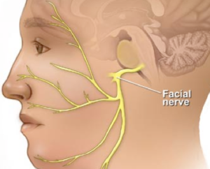
The localization of HHV-6 in the facial nerve ganglia and trigeminal ganglia suggests that severe primary infection and reactivation may result in damage or dysfunction in these areas. Indeed, herpesviruses, including HHV-6, have been implicated in cases of abnormal cranial nerve function. HHV-6 has been implicated in facial palsy (Kanerva 2013, Pitkäranta 2004), trochlear (fourth) nerve palsy (Papageorgiou 2014, Thierfelder 1997), and optic neuritis (Ogata 2011, Hino 2016, Moschettini 2006, Mechai 2007).
HHV-6 has previously been detected in the trigeminal, geniculate, vestibular, and dorsal root ganglia (Hüfner 2007). It is hypothesized that HHV-6 enters the central nervous system through the nasal cavity and thereby the olfactory pathway, in which the virus is detected frequently. Among a subset of adults who died after courses of unspecified encephalopathy, ultrastructural changes in the olfactory bulb/tract may be associated with HHV-6-induced dysfunction.
The presence of HHV-6 in these ganglia reinforces the neurotropism of the virus and suggests that these nerves may serve as important reservoirs of latent HHV-6.
Find the full paper here: Ptaszyńska-Sarosiek 2019.