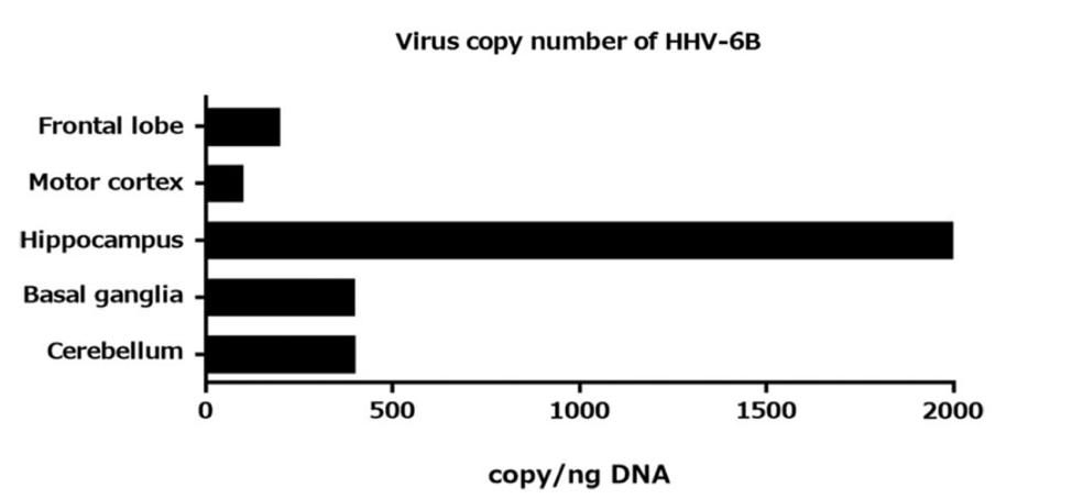
The autopsy of an infant with HHV-6B encephalitis showed a viral load 4-5 fold higher in the hippocampus compared to other parts of the brain. Neurons, oligodendrocytes and vascular endothelial cells were infected, but not astrocytes or microglia.
 The infant, who died three days after developing a seizure, had massive brain edema. Microscopic examination showed swollen astrocytes and ballooned oligodendrocytes in the frontal white matter, along with neuronal cell death and microglial infiltration in the frontal cortex. Of interest, the hippocampus had neither neuronal loss nor reactive glial response.
The authors speculate that reactivation of silent and abundant HHV-6B infection in the hippocampus might be associated with hippocampus-oriented disorders such as temporal lobe epilepsy.
For more information, read the full paper: Miyahara 2018