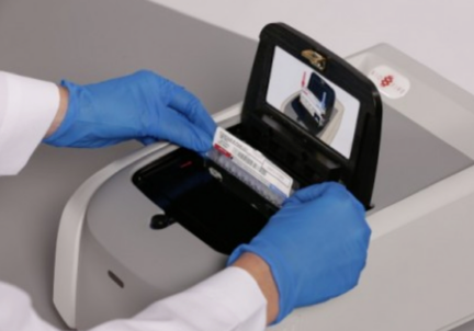
This multiplex qualitative test for HHV-6 and other pathogens in cerebrospinal fluid allow physicians to diagnose HHV-6 as a cause of encephalitis quickly, especially when interpreted in conjunction with imaging, blood tests and determination of ciHHV-6 status.
A chart review of 25 patients whose CSF tested positive for HHV-6 using the FilmArray Meningitis/Encephalitis (FA-ME) panel aimed to evaluate the clinical significance of a positive result via this testing modality. Often, it is difficult to determine whether HHV-6 is active and causing or contributing to symptoms, is inactive yet present in the specimen tested, or whether it is reactivating opportunistically with no effects on the clinical course. Use of the assay has resulted in skepticism over its utility in the past because of the difficulties associated with determining the clinical relevance of a positive result.
In taking a broader view and analyzing the clinical course, iciHHV-6 status, coinfections, treatments, and other factors, it was concluded that HHV-6 positivity on the FA-ME panel led to appropriate initiation and discontinuation of drugs, shorter time to determining the cause of illness, and when results are considered alongside other diagnostic tools, including MRI and blood testing, that the assay is very useful in the clinical setting.
A total of 1,005 pediatric patients with signs and symptoms of meningitis or encephalitis underwent FA-ME testing, and 25 (2.5%) were HHV-6-positive, two of whom were also positive for another pathogen (enterovirus and Streptococcus pneumoniae) via the FA-ME assay. However, only 5 were diagnosed with HHV-6 meningoencephalitis (4) or meningitis (1), while another 4 were found to have probable HHV-6 meningoencephalitis/meningitis. The 5 who received a diagnosis were between 0.76 and 12.87 years old, and all 4 of the children with probable HHV-6 meningoencephalitis/meningitis were under a year old. Only one of the 9 was not previously healthy, having received a stem cell transplant prior to his meningoencephalitis. Ganciclovir, foscarnet, or a combination of ganciclovir and valganciclovir were administered to the 4 children with HHV-6 meningoencephalitis and 2 additional patients, but the child with meningitis was not given antiviral medication. One of the immunocompetent patients with meningoencephalitis, as well as the patient who received a stem cell transplant, died.
The children with probable HHV-6 meningoencephalitis/meningitis had self-limited bulging fontanelles and fever, but in the two patients who underwent radiological imaging, only mild prominence of extra-axial spaces and small extra-axial fluid collections were noted, whereas all 5 children with diagnosed HHV-6 meningoencephalitis/meningitis had imaging abnormalities.
11 children had presumptive primary HHV-6 infection without clinically significant central nervous system disease. Four of them also had positive urine cultures, and another had a nasopharyngeal swab positive for enterovirus/rhinovirus. Among the patients with diagnosed HHV-6 meningoencephalitis/meningitis, EBV was detected in the CSF of one but was considered to be episomal, another had several other viruses detected in the blood and nasopharyngeal swab sample, resulting in a diagnosis of adenovirus viremia along with the meningoencephalitis, and a third had norovirus in a diarrheal stool specimen.
Ganciclovir/valganciclovir was given to one patient without diagnosed HHV-6 meningoencephalitis/meningitis who was considered to have probable reactivation (HHV-6 was untyped) and CNS demyelination. Imaging abnormalities were found in this 17-year-old patient, she exhibited weakness, sensory loss, fever, abnormal gait, and headache, and no other pathogens were identified. While it was unclear what the role of the virus was in this patient, HHV-6 has been implicated in demyelinating diseases.
Of 18 patients tested for iciHHV-6 by ddPCR, two were confirmed to have iciHHV-6B and one had iciHHV-6A. The rest of the 11 patients whose HHV-6 was typed had HHV-6B. None of the iciHHV-6-positive children were diagnosed with nor treated for HHV-6 meningoencephalitis/meningitis. Two of three had sepsis: One, a one-year-old girl who did not survive, was on chemotherapy for brain cancer and had several other pathogens detected in different compartments. The second was under a month old and presented with fever, jaundice, congestion, and cough. The third iciHHV-6 positive patient was also under a month old, exhibited pleocytosis, and was admitted with a fever.
Whole blood HHV-6 PCR was performed alongside the FA-ME assay in 11 patients and was positive in the majority (9). Interestingly, one of the patients diagnosed with HHV-6 meningoencephalitis was strongly positive for the virus in the CSF but was consistently negative in the blood, which the authors note suggests that viremia may not always accompany HHV-6-associated meningoencephalitis/meningitis. The rest of the meningoencephalitis/meningitis patients whose whole blood was tested (3) were positive.
While HHV-6 is often considered a pathogen limited to the setting of severe immunosuppression, only 3 of the patients in this cohort of 25 had underlying issues, and only one was a stem cell transplant recipient. In most of these HHV-6-positive cases, the virus appeared to contribute or cause the presenting symptoms, although it was only associated with meningoencephalitis/meningitis in 20% of cases. HHV-6 positivity via the FA-ME assay resulted in withdrawal of empirical acyclovir therapy in all patients who initially received it, and incidental detection of ciHHV-6 did not lead to overuse of antiviral medication. Together, these findings indicate that HHV-6 positivity on the FA-ME panel provides useful, clinically relevant insight into the etiology of symptoms as well as allows for appropriate discontinuation of acyclovir and antibiotics.
Find the full paper here: Pandey 2020.

