Surprisingly, the damage from HHV-6B infection often occurred in the absence of obvious neurological symptoms. Patients without HHV-6 reactivation had no reduction in volume.
An earlier study led by Danielle Zerr of University of Washington found that HHV-6B infection is associated with mild cognitive impairment in hematopoietic stem cell transplants (HSCT) (Zerr 2011). Tetsushi Yoshikawa of Fujita Health University designed a study to further elucidate this correlation in pediatric patients. 107 patients undergoing HSCT had weekly monitoring for HHV-6 infection by both viral isolation and real-time PCR of whole blood. Of the 107 patients, 20 had MRIs before and after HSCT that were technically adequate for analyzing hippocampal volume. Of these 20, 8 developed HHV-6B reactivation (as judged by either viral isolation or high viral load) and could be compared to the 12 who did not.
Of the 8 patients with HHV-6B reactivation, 2 had encephalopathy; however, the remaining 6 had no obvious clinical deficits on neurological examination. Of the 12 without HHV-6B reactivation, none had encephalopathy or obvious deficits on neurological examination.
Hippocampal volumetric measurements were performed using coronal T2-weighted slices. The hippocampal volume ratio was calculated by dividing the hippocampal volume after transplantation by the volume before transplantation (<1.00 indicates volume loss).
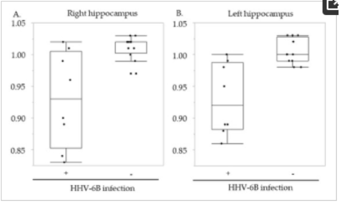
The hippocampal volume ratio before and after transplantation. (A) Right hippocampus; (B) Left hippocampus. The vertical axes indicate the hippocampal ratio of the hippocampal volume from before to after hematopoietic stem cell transplantation recipients with (+) and without (−) human herpesvirus 6B (HHV-6B) infection. - Hippocampal Atrophy in Pediatric Transplant Recipients with Human Herpesvirus 6B (Miyake et al. Apr 2021)
In the right hippocampus, the median volume ratio was 0.93 in patients with HHV-6B infection and 1.02 in patients without HHV-6B infection (p=0.007). In the left hippocampus, the same ratios were virtually identical: 0.92 and 1.00, respectively (p=0.003).
Because only 20 of the 107 patients undergoing HSCT had adequate MRI volumetric studies, it is not clear how representative these 20 patients studied were of the larger group of 107 people undergoing HSCT. This study suggests that reactivated infection with HHV-6B during HSCT may be associated with hippocampal volume loss, even in children without evidence of neurological deficits on standard neurological examination. However, earlier work by Zerr and others has found that subtle cognitive deficits may be more difficult to detect in pediatric patients, requiring more sensitive neuropsychological testing.
Read the full article: Miyake 2021