Levels of mRNA transcripts indicating active infection strengthen case for pathogenic role.
“Idiopathic pneumonia syndrome” (IPS) occurring after allogeneic hematopoietic cell transplant (HCT) refers to a pneumonia in which the “usual” infectious agents are not present. It has a high mortality rate.
Many past studies, the first more than 30 years ago, have found that HHV-6B DNA is found in the bronchioalveolar lavage fluid, or BALF (washings obtained using a bronchoscope to collect fluid and cells from the lungs) of people with IPS. Furthermore, past studies have found a higher mortality rate in people with IPS who have HHV-6B DNA in the BALF (Hill 2019). HHV-6B gene expression in lung tissue has been documented using RNA in situ hybridization. HHV-6B has been shown capable of infecting bronchial gland cells (Krueger 1990). A mouse model infected with murine roseolovirus (a homolog of HHV-6) reactivates in the lung and produces an IPS-like phenotype (Zhou 2019).
Despite this substantial past evidence indicating a pathogenic role for HHV-6B in IPS, many have remained skeptical. Given that most people have white blood cells that are chronically infected with HHV-6B—and can enter BALF—simply finding viral DNA in BALF is not strong evidence that HHV-6B is actively replicating and acting as a pulmonary pathogen.
A team from the University of Washington and Fred Hutchinson Cancer Center, including Joshua Hill and Michael Boeckh, conducted a prospective multicenter study of 116 patients with IPS after allogeneic HCT. The BALF and plasma were tested for the presence and levels of HHV-6B DNA and for two HHV-6B mRNA transcripts associated with lytic infection—U38 (a late DNA polymerase gene) and U90 (an immediate early gene).
HHV-6B DNA was found in 37% of the BALF specimens, but only in 15% of plasma samples. Interestingly, HHV-6B mRNA transcripts were found in an even higher number (49%) of specimens, suggesting that measuring DNA in BALF or plasma may be less sensitive than measuring mRNA in BALF, as well as less specific for active infection. Higher HHV-6B DNA viral loads in either BALF or plasma predicted HHV-6B mRNA detection. Those same high viral load measurements also predicted increased overall mortality and death due to pulmonary failure.
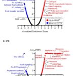
To find other evidence that lytic infection with HHV-6B was, in fact, eliciting an immune response, the team also evaluated host gene expression signatures. They found distinctive patterns of upregulation and downregulation of various gene pathways in people with evidence of lytic infection. The figure below displays a volcano plot of results from pathway analysis using Gene Set Enrichment Analysis (GSEA) to define and compare transcriptional programs in people with other viral pneumonias and in those with IPS (many of whom had evidence of active replication of HHV-6B).
As pointed out by Moore et al (Victoria 2020) most past studies of infections in IPS have either failed to test for HHV-6, tested only plasma and not BAL fluid, or used a qualitative PCR which cannot differentiate low-level positivity from active infection.
The findings of this study provide the strongest evidence yet that HHV-6B is an important cause of IPS following allogeneic HCT. The investigators demonstrate that the pathogenic role of the virus is most certain when viral loads in BALF are ≥2.8 log10 copies/mL. Since IPS patients are often given high dose steroids which exacerbate HHV-6 infection, the findings underline the urgency of clinical studies of both diagnosis and treatment of HHV-6B in people with IPS following allogeneic HCT.
Read the full article: Hill 2023