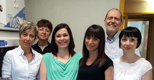
 A new study reported that HHV-6A infects the lining of the uterus in 43% of women with unexplained infertility but cannot be found in the uterine lining of fertile women. Furthermore, the cytokine and the natural killer cell profiles were very different in patients with the infection. HHV-6A was found only in uterine endothelial cells, and not in the blood.
A team led by Roberta Rizzo, Roberto Marci, and Dario DiLuca of the University of Ferrara tested endometrial biopsies from 30 women with unexplained primary infertility as well as 36 fertile women. HHV-6 DNA was found in the endometrial samples from 13 of 30 infertile women (43%), but none of the fertile women. Surprisingly, all of the virus was HHV-6A, not HHV-6B, the virus that reactivates in immunocompromised patients and causes roseola in infants. Over 97% of HHV-6 reactivation in transplant patients is typed as HHV-6B.
Of interest, the investigators found a strong correlation between the level of estradiol and presence of an HHV-6A infection (p=0.02). They also found that the virus was active only during the secretory phase of the menstrual cycle, when estradiol levels were highest. Estradiol has also been shown to cause HSV1 reactivation (Miguel 2010), and steroids cause HHV-6 to reactivate disproportionately in patients with DIHS/DRESS (Ishida 2014).
Cytokine and natural killer (NK) cell levels were significantly different between women who were HHV-6A positive compared to controls and infertile women who were HHV-6A negative. While the Th2 cytokine IL-10 was higher in the HHV-6A positive group, the Th1 cytokine IFN-gamma was lower than in controls or HHV-6A negative women, causing an increase in the Th1/TH2 ratio, a condition common in female infertility. The authors note that HHV-6 is known to increase IL-10 expression by monocytes and reduce IFN-gamma by T lymphocytes.
In addition, levels of a specific subset of uterine NK cells were significantly lower in HHV-6A+ individuals than in the controls, and uterine NK cells from the HHV-6A infected group showed higher activation in response to the virus than the cells collected from controls. The authors suggest that these findings are consistent with an abnormal, persistent immune response toward HHV-6A antigens in this subgroup of women with unexplained infertility.
The HHV-6A DNA was found only in the endometrial epithelial cells, and not the uterine stromal cells. No HHV-6A was found in the peripheral blood, and latent HHV-6B was found in equal amounts in the blood of both patients and controls. As a result, the condition can be diagnosed only by testing a uterine biopsy.
A Japanese study showed that HHV-6 IgG and IgM titers were significantly higher in patients with spontaneous abortions at 6-12 weeks compared to controls, and 8% were IgM positive. HHV-6 antigens were in the villous tissues (Ando 1992). Several other studies have also found HHV-6 DNA in fetal tissue after miscarriage (Drago 2008, Drago 2014, Revest 2011). Past studies have also found altered cytokine levels in the uterine tissue of infertile women (Ozkan 2014).
How is HHV-6A acquired?
Very little is known about the prevalence of HHV-6A or how it is acquired. Although 95% of us shed significant amounts of HHV-6B in the saliva, HHV-6A is found in such low amounts it cannot generally be detected in saliva by standard testing (Aberle 1996). A study at the NINDS using more sensitive techniques found low levels of HHV-6A in the saliva of 12% of healthy controls compared to 20% of patients with MS (Leibovitch 2014). HHV-6 remains latent for life, and typically only reactivates in patients who have an immune defect or who are taking immunosuppressive drugs.
HHV-6 in the semen?
HHV-6 has also been found in 13.5% of semen donations, causing some speculation as to whether the uterine infections might be sexually transmitted (Kasperson 2012). Much higher rates of viral infections have been reported in men with infertility, but no study has characterized the virus found in men with infertility to determine the prevalence of HHV-6A. The virus found in healthy controls appears to be predominantly HHV-6B.
HHV-6A has also been linked to Hashimoto’s thyroiditis (Caselli 2012) and has been implicated as a trigger in a subset of multiple sclerosis patients. Approximately 20% of oligoclonal bands found in the spinal fluid of MS patients are HHV-6 specific (Virtanen 2014) and have been found to react to an HHV-6A lytic protein (Alenda 2014). HHV-6A and HHV-6B have different receptors and significantly different cell tropism (Ablashi 2014). For example, HHV-6A but not HHV-6B can infect natural killer cells.
NOTES FOR PATIENTS AND FERTILITY CLINICIANS:
Testing:
Commercial labs that can test uterine biopsies for HHV-6 include Coppe Laboratories and ViracorIBT in the US as well as IKDT in Germany. (See “How to get a uterine biopsy tested for HHV-6A“)
ABOUT THE AUTHORS:
Roberta Rizzo, PhD coordinated the study that was funded through the “Allesandro Liberati Young Scientists Award”. Dario DiLuca, PhD is a professor in the Department of Medical Sciences, and Roberto Marci, MD, is a professor of Morphology, Surgery, and Experimental Medicine.

Elisabetta Caselli, Antonella Rotola, Valentina Gentili, Daria Bortolotti, Dario Di Luca, Roberta Rizzo

