NINDS investigators tested the saliva of 115 children with acute seizures and found that they had elevated inflammatory cytokines compared to healthy controls and children with fever. One of those cytokines, Il-1β, correlated with HHV-6 saliva viral load.
The NINDS group, led by Luca Bartolini, MD, and Steve Jacobson, PhD, established that elevations of both IL-1β and IL-8 were much higher in the saliva of infants with seizures – demonstrating that these cytokines are not influenced by the fever itself, but more likely an underlying infection. Children with seizures are not routinely tested for HHV-6 and current treatment with antiepileptic drugs is symptomatic. Drugs targeting potential mechanisms, such as antiviral or anti-inflammatory treatments in epilepsy are still considered experimental.
Bartolini and colleagues tested for EBV and HHV-6 in the saliva of 115 children with acute seizures, including new onset unprovoked seizures, breakthrough seizures in chronic epilepsy, febrile seizures, and newly diagnosed epilepsy in addition to 51 children with fever alone and 46 older healthy children. There was no significant difference in prevalence between the cases and control groups. However, differences in prevalence are not generally expected with a ubiquitous virus. Also, small children must be carefully matched in age for a prevalence comparison since many have not had their primary infections. The healthy control children used in this study were older, while those with febrile seizures were mostly infants.
Past studies have shown that elevated levels of IL-1β, IL-8, and TNF are found in the serum and CSF of patients with epilepsy (Gallentine 2017), and HHV-6 has been shown to induce dysregulation of glutamate homeostasis in astrocytes and contribute to seizure formation (Fotheringham 2007).
The largest study of HHV-6 in febrile seizures was performed by Carolyn Hall and colleagues and was published in 2004 in the NEJM. In their cohort, they found that 17% of 146 children presenting with seizures had HHV-6B primary infection (Hall 2004). Since they did not include HHV-6 reactivations in this count, the true number involving HHV-6 infections was presumably significantly higher.
A comprehensive study of HHV-6B in febrile status epilepticus found that 32% of children were positive for HHV-6B. Of those, 30% that were reactivations, and 70% were primary infections (Epstein 2012). In the Epstein study, they were not measuring prevalence of HHV-6 (as with the NINDS saliva study), but rather active infection as determined by mRNA, IgM antibodies or a four-fold increase in IgG antibodies.

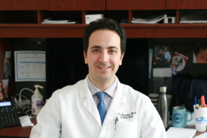
Luca Bartolini, PhD, Clinical Epilepsy Section and Division of Neuroimmunology and Neurovirology, NINDS, NIH
Although blood was not evaluated in this NINDS study, saliva may be a more sensitive method of detecting HHV-6 reactivation in children. One study comparing blood and saliva of adult leukemia patients in 2015 showed that HHV-6 viral loads are much higher in saliva than in the whole blood and that high levels in the blood generally correlated with high levels in the saliva. Furthermore, they found that an increase in the HHV-6 DNA in saliva preceded an increase in the blood. However, the majority of leukemia patients were positive for HHV-6 or CMV in saliva, but negative in the blood (Nefzi 2015).
The authors plan future larger studies to study viral DNA presence in saliva and simultaneous blood and cytokine expression over time. Hopefully these studies will include testing of HHV-6B viral load as well.
For more information, read the full paper: Bartolini 2018.

