HHV-6 is rarely identified as the cause of liver dysfunction in immunocompetent children, in part because HHV-6 is not included in routine testing, and HHV-6 infections can be highly localized to the liver. In this case, an alert team in Arizona identified HHV-6 by needle biopsy, but the diagnosis was too late to prevent a transplant.
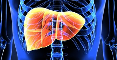
A previously healthy and immunocompetent 11-year-old boy presented with a history of nausea, non-bilious, non-bloody emesis, and fatigue. He was diagnosed with acute viral gastroenteritis and soon developed a nonspecific generalized rash on his body. Within that same week, he developed significant jaundice, anorexia, headaches, and increased fatigue. He was transferred to a different hospital, where his initial lab assessment revealed elevated liver enzymes (AST 2553 U/L, ALT 1966 U/L). Hepatitis panel results were negative and liver ultrasound was normal so the patient was discharged.
The next day, he developed a 102ºF fever and was transferred to another facility. Tests revealed worsening liver function which prompted a transfer to Phoenix Children’s Hospital. Follow-up tests (AST 3585 U/L, ALT 2278 U/L) suggested that the patient was experiencing acute liver failure (ALF). An extensive work-up for underlying etiologies ruled out Wilson disease, autoimmune hepatitis, α-1 antitrypsin deficiency, vascular abnormalities, Coccidioides, human immunodeficiency virus (HIV), HSV I and II, Mycobacterium, respiratory syncytial virus (RSV), CMV, EBV, parvovirus B19, and influenza A and B. Additionally, an acute hepatitis serology panel ruled out Hepatitis A, B, C, and E. The only positive findings initially were rhino/enterovirus detected in a nasopharyngeal swab and evidence of previous EBV exposure.
Of note, HHV-6 was not included in any initial testing by the multiple institutions in this case report, highlighting the lack of awareness of HHV-6 as a pathogenic virus, particularly outside of the infectious disease and transplant community. As the authors noted in their discussion, “many pediatric patients are not fully tested for viral etiologies, especially HHV-6”. They think that the lack of broad viral screening in clinical practice is counterintuitive given that the etiologies of 40-50% of fulminant hepatitis cases are idiopathic. The timeliness of HHV-6 detection is critical to successful therapy (Phan 2017).
A liver biopsy consisting of several needle cores obtained from the patient demonstrated acute hepatitis characterized by submassive hepatic necrosis and architectural collapse. Immunohistochemical staining (IHC) and qPCR for HSV I and II, CMV, adenovirus, EBV, and hepatitis B, yielded negative results. However, qPCR for HHV-6 was positive in the needle biopsy (1250 copies/mL). Interestingly, IHC analysis for HHV-6 was negative and HHV-6 was never detected in the plasma of this patient despite significant infection in the liver. The localization of HHV-6 infection in the liver with no detectable viremia has been reported in the literature (Buyse 2013, Phan 2017). Furthermore, this phenomenon has been reported in other organs as well, including the lungs (Seo 2015), heart (Leveque 2011), and CNS (Caserta 2013).
The patient was treated with prophylactic antibiotics, antifungals, and acyclovir. Acyclovir was replaced with valganciclovir after detection of HHV-6 via qPCR. Despite treatment, the patient’s liver function declined rapidly and he developed worsening hepatic encephalopathy. He underwent a successful ABO-compatible liver transplantation within 3 days of admission.
Liver explant revealed progressive degenerative changes with massive hepatic necrosis (>99%) compared to the initial biopsy (65%) obtained less than 1 week prior. Bile ductular proliferation was conspicuous, as were moderate portal lymphocytic infiltrate and venulitis. A 1.5-cm wedge of the explant was retested for HHV-6 (37,689 copies/mL). This increase in viral load was ascribed to specimen size and/or location.
Further IHC testing revealed HHV-6B in the ductular epithelium. No other evidence of HHV-6 or other etiologic agents were identified in the liver or plasma, reinforcing the hypothesis that the ALF was caused by a localized, active HHV-6 infection.
The post-transplant course of treatment was complicated by pleural effusions, ascites, and pancytopenia. The pleural effusion was also positive for HHV-6 (5,000 copies/mL). Primary HHV-6 infection in a pediatric patient after liver transplantation associated with HHV-6 detection in pleural effusions has been successfully treated in the past with antivirals and reduction in immunosuppression (Dohna-Schwake 2011).
Bone marrow biopsies were analyzed for HHV-6: the first was negative but the second was positive for HHV-6. However, no HHV-6 positive cells were identified by IHC, thus the authors ruled out direct myelosuppression by viral infection. They attributed their bone marrow findings to aplastic anemia coinciding with acute hepatitis. The patient underwent platelet transfusion and was treated with granulocyte colony-stimulating factor which resulted in increased cell counts. The patient is still being treated with prophylactic and post-transplant therapeutic protocols and he continues to test negative for HHV-6.
This case report exemplifies the need to raise awareness of the pathogenicity of HHV-6 in the medical community as well as the need for more funding for HHV-6 research. To date, we still do not completely understand how HHV-6 causes liver failure. Although HHV-6B is primarily a CD4-lymphotropic virus, it can infect a variety of cell types, from monocytes to hepatocytes to ductal epithelia, as shown in this case report. Most of our knowledge of clinical HHV-6 infections stem from hematopoietic stem cell transplant (HCT) studies, and it is highly likely that HHV-6 uses different mechanisms to cause disease in the HCT setting compared to that of solid organ transplantation. Advances in our understanding of HHV-6 biology and the immune response to HHV-6 infection will catalyze the development of more specific and less toxic antiviral therapies and protocols.
Read the full study: Szewc 2018