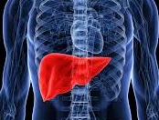
A review published in Liver International summarizes and highlights findings from past studies which demonstrate that qPCR in the blood is insufficient for identifying hepatic HHV-6 infection/reactivation in liver transplant patients. This is because HHV-6 reactivation can be restricted to the infected organ with no evidence of active infection in the blood. For this reason, the authors emphasize that only liver biopsy (confirming histopathological effects of HHV-6 in the tissue itself) should be considered reliable for diagnosis of liver disease.
HHV-6 infection/reactivation in liver transplant patients is rarely explored or diagnosed, but in reported cases of significant tissue-invasive infections, patients have been treated successfully with antivirals. Clinical symptoms of infection include fever, hepatitis, encephalitis, myelosuppression, and higher rates of graft dysfunction as well as indirect effects including increased risks of mortality, CMV disease, hepatitis C progression, and greater fibrosis scores.
In a detailed analysis of the published literature on HHV-6 in liver transplant patients, the group reports that the frequency of HHV-6 reactivation after liver transplantation is approximately the same in plasma (33%) and PBMCs (34%) overall. However, reactivation rates are influenced by the administration of CMV prophylaxis, as just 50 (11%) of the 455 patients receiving CMV prophylaxis reactivated HHV-6 compared with 167 (39%) of the 433 patients receiving acyclovir or no prophylaxis.
The authors also note that inherited chromosomally integrated HHV-6 (ciHHV-6), in either the recipient or the donor organ, may create confusion surrounding the diagnosis of systemic HHV-6 infection/reactivation in affected patients. Liver transplant recipients with inherited ciHHV-6 may have an increased risk of opportunistic infection and graft rejection (Lee 2012), something that has also been observed recently in the setting of HCT (Hill 2017b).
Although HHV-6 infection is currently diagnosed by quantifying viral DNA in plasma or blood, biopsy to demonstrate histopathologic effects of HHV-6 remains the gold standard for diagnosis of end-organ disease. HHV-6 reactivation may be restricted to the infected organ with no evidence of active infection in the blood. A study (Buyse 2013) found that HHV-6 DNA was detectable in 10 of 26 (38.5%) liver biopsy specimens diagnosed with graft acute hepatitis of undetermined origin. In 4 (40%) of the 10 patients, confluent periportal necrosis was associated with high tissue HHV-6 viral load on liver biopsy. In contrast, periportal necrosis was absent in graft hepatitis not documented to be associated with HHV-6. Hence, the presence of confluent periportal necrosis with high intragraft viral load may indicate HHV-6-induced hepatitis. Notably, only 28.6% of patients with HHV-6-positive graft hepatitis had elevated levels of HHV-6 in the PBMCs associated with active infection. The authors concluded that in over two thirds of cases, the reactivation of HHV-6 would have been missed if the testing had been performed only on the peripheral blood. As a result, HHV-6-associated liver disease cannot be ruled out in the absence of a biopsy even when there is no detectable viremia. Within the same patient groups, the rates of HHV-6 detection have been approximately 56.8% in tissue biopsies (range 20% to 100%) vs. only 17.6% in blood samples (range 10% to 83.3%) (Phan 2017). If HHV-6-induced liver dysfunction is suspected, liver tissue will be more revealing than plasma or serum, although both should still be tested for HHV-6. The localization of HHV-6 in target organs without detection in the blood can be found in other organs as well, especially during persistent infections: HHV-6 DNA can be found in lung biopsies but not the plasma of patients with “idiopathic” pneumonia syndrome after lung transplantation (Seo 2015) and high levels of DNA can be found in biopsy tissues of patients with persistent HHV-6 myocarditis, in spite of little or no DNA in the plasma (Leveque 2011).
HHV-6 is often reactivated from latency during periods of intense immunosuppression, although sporadic cases of HHV-6 reactivation have been reported even in the immunocompetent (Leveque 2011, De Simone 2013, Cacheux 2005, Fujita 2015).
Read the full paper: Phan 2017